• Hello
  • Notes
  • FAQs
    • My Amazon Shop
    • MY LTK
    • MightyHoop / HoopMaster
    • PhoneSoap
  • KIND WORDS
Menu

THE NURSE NATALIE

  • Hello
  • Notes
  • FAQs
  • SHOP
    • My Amazon Shop
    • MY LTK
    • MightyHoop / HoopMaster
    • PhoneSoap
  • KIND WORDS
Screen Shot 2020-05-24 at 9.26.15 AM.png

Conjugated vs. Unconjugated Bilirubin

May 24, 2020

Hold up, wait a minute! You mean to tell me there’s TWO types of bilirubin? Mind = blown! 

In one of my previous posts, we discussed bilirubin and why it’s important to monitor. Today, we are going to talk about the two different forms.

So... what’s the difference?

In short, one is considered “normal” and shows up after a couple days of life. The other is “abnormal” and usually appears within the first few days of life.

Unconjugated Bilirubin

  • Also known as “indirect” bilirubin or the “normal” type.

  • This type is unable to be conjugated by the liver and re-enters enterohepatic circulation in two different ways:

  1. Bound to albumin

  2. Unable to bind to albumin and remains free in the plasma to potentially move into tissues

Conjugated Bilirubin

  • Also known as “direct” bilirubin or the “abnormal” type

  • It is processed by the liver and is ready to be excreted through stool and urine

What causes unconjugated bili to increase?

  • Polycythemia

  • Hemolysis of RBCs d/t Rh incompatibility or ABO incompatibility between mom and baby

  • Traumatic birth resulting in hemorrhage, bruising, cephalohematoma

  • Preterm birth

  • Inability to pass stool or make urine (excretion disorder)

    • May be from Hirschprung’s disease, imperforate anus, intestinal atresia, meconium ileus, etc.

  • Immature liver

  • Infection, sepsis

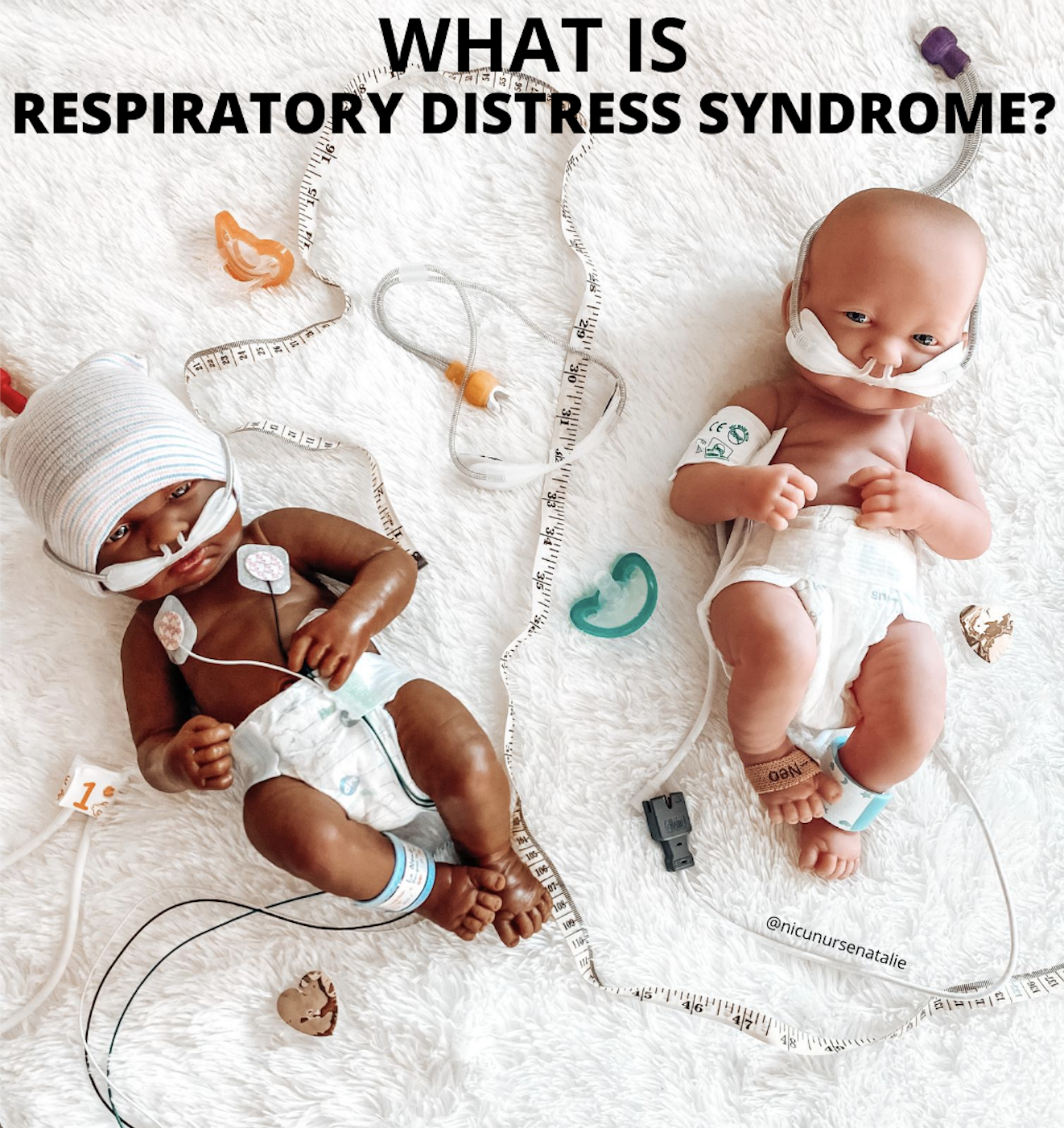
  • IDM, RDS—Since conjugation requires oxygen and glucose, a lack of these nutrients will cause bili to increase

  • Some medications compete with bilirubin for albumin binding sites (e.g. NSAIDs, steroids, lipids, etc.)

What causes conjugated bili to increase?

  • Chronic, long-term TPN and Intralipid use—This causes damage to the liver and biliary tree and affects bilirubin excretion

  • Biliary obstruction/atresia/cholestasis—If bile cannot be excreted from the liver, bilirubin cannot be excreted and will be reabsorbed.

  • In-born error of metabolism (e.g. galactosemia)

How do you treat unconjugated bili?

  • Phototherapy—Uses UV light to breakdown unconjugated bili into a water-soluble form that can be excreted

  • Hydrate with IV fluids

  • Feed the baby! Unconjugated bili is eliminated via stool and urine!

  • Exchange transfusion in critical cases

How do you treat conjugated bili?

  • Get the baby off TPN!

    • If not, remove the copper & manganese from the bag—An accumulation of these can be toxic if the liver is not functioning properly.

  • Get the baby off Intralipids!

    • If not, switch the baby to SMOF lipids. This fat emulsion reduces inflammation, oxidative stress, and/or cholestasis.

  • Phenobarbital. This med increases bile acid synthesis and bile secretion

  • Actigal/Ursodiol. This med increases bile acid secretion and bili excretion

  • Pregestimil formula. This type of formula is extremely hydrolyzed and specific to infants who suffer from fat malabsorption.

  • AquaDEKs (ADEKs) Multivitamin. This med is high in the fat-soluble vitamins that these infants may be deprived of.

Comment
pyloric stenosis.gif

Pyloric Stenosis

May 22, 2020

What is Pyloric Stenosis?

Pyloric stenosis is a problem that affects many newborns and causes forceful vomiting that can lead to dehydration. These babies require a high level of care and will be admitted to the NICU. It is the second most common problem requiring surgery in newborns.

The lower portion of the stomach that connects to the small intestine is known as the pylorus. In pyloric stenosis, the muscles in this part of the stomach enlarge, narrowing the opening of the pylorus and eventually preventing food from moving from the stomach down to the intestine. Normally, this muscular valve (pylorus) holds food in the stomach until it is ready for the next stage in the digestive process. In pyloric stenosis, the pylorus muscles thicken and become abnormally large, blocking food from reaching the small intestine.

These babies will experience persistent, projectile vomit, ejecting breast milk or formula up to several feet away. Vomiting might be mild at first and gradually become more severe as the pylorus opening narrows. The vomit may sometimes contain blood.

Which Babies are at Highest Risk?

Pyloric stenosis can be inherited; several members of a family may have had this problem during infancy. In addition, pyloric stenosis is four times more common in males than females.

Why is Pyloric Stenosis Concerning?

The most serious problem that can result from pyloric stenosis is dehydration. When a baby vomits regularly, he or she will not get enough fluids to meet his or her nutritional needs. Babies have smaller bodies than adults and cannot tolerate losing fluid as easily as we can. Your baby might cry without tears or become lethargic. You might find yourself changing fewer wet diapers in a day or diapers that are completely dry.

Electrolyte imbalance is another problem that pyloric stenosis causes. Minerals that the body needs to stay healthy, such as potassium and sodium, are lost from the frequent vomiting. Babies who are deficient in key electrolytes can become sick very quickly. Lack of vitamins and nutrients can lead to malnutrition, and ultimately—weight loss. A baby who vomits most of (or all of) his or her feedings will not have adequate nutrition to gain weight and stay healthy. This can lead to failure to grow, develop, and thrive.

Lastly, stomach irritation is another complication that can occur. Repeated vomiting can irritate your baby's stomach and may cause mild bleeding or ulcers.

What are the Symptoms of Pyloric Stenosis?

The most common symptoms noted in a baby with pyloric stenosis is forceful, projectile vomiting. This kind of vomiting is different from a "wet burp" or an occasional spit up that a baby may have at the end of a feeding. Large amounts of breast milk or formula are vomited and may project several feet across the room. The milk is often curdled in appearance—as the milk remains in the stomach and does not move forward to the small intestine, the stomach acid "digests" it.

In addition to the complications previously discussed, other symptoms may include:

  • Persistent, constant state of hunger

  • Eats or nurses eagerly

  • Lack of energy

  • Fewer bowel movements (constipation)

  • Frequent, mucous stools 

  • Stomach contractions

How is Pyloric Stenosis Diagnosed?

In addition to a complete and thorough medical history and physical examination, a barium swallow study/upper GI series is typically warranted. This is a diagnostic test that examines the organs of the upper part of the digestive system: the esophagus, stomach, and duodenum (the first section of the small intestine). A fluid called barium (a metallic, chalky liquid) is swallowed and used to coat the inside of your baby’s organs so that they illuminate on X-ray. A series of X-rays are then taken to evaluate the digestive organs and to track the progression of the dye through the tract.

How is Pyloric Stenosis Treated?

Pyloric stenosis MUST be repaired with an operation and will NOT resolve on its own. A pyloromyotomy is performed, which is an operation to loosen the tight muscle causing the blockage between the stomach and small intestine. The surgeon makes an incision in the muscle fibers of the enlarged pyloric muscle. He will cut through the wall of the pylorus to widen the opening into the intestine. This loosens the tight muscle causing the blockage. The lining of the pylorus will then bulge through the incision, opening a channel from the stomach to the small intestine. After surgery, your baby may still vomit, but this will resolve after a few days. Dehydration and electrolyte imbalances will be treated via IV fluids. In addition, he or she will remain on antiemetic medications such as Pepcid, Reglan, Prevacid, etc.

img_main.gif
ds00815_im00225_r7_pyloric_stenosis_thu_jpg.png.jpeg
ds00815_im04773_mcdc7_pyloric_stenosisthu_jpg.png.jpeg
Comment
Adjustments.jpeg

I Got My Antibodies Tested

May 19, 2020

One of my favorite things to do during my spare time is read all of the crazy conspiracy theories about COVID-19. Some of the hypotheses are so bizarre and far-fetched, yet super interesting and entertaining. Some people are calling this whole thing a big, fat hoax and rumoring that it’s just a political stunt because it’s election year. Mind = blown! I also love observing all of the “Chicken Littles” in the world—you know who you are—running around screaming the fable’s central phase: “The sky is falling!” There are SO many people out there accustomed to being unreasonably afraid and overly paranoid, or those trying to incite fear in everyone around them. People are living on both sides of the spectrum.

When the Central Valley issued its first shelter-in-place order on March 18th, my fiancé and I started preparing to spend the next few weeks in quarantine, isolated from the rest of the world.

With everything shut down and nothing to do, we spent all of our time mindlessly watching the news for hours on end. It was so fascinating to me hearing how so many people who tested positive for the virus did not show the “classic” signs and symptoms. That is—fever, shortness of breath, loss of smell, etc. Some reported a wide range of symptoms, while others didn’t show any. Did that mean it was possible to catch coronavirus and not know it?

With that thought in mind, I acted on my curiosity and decided to take the SARS-CoV-2 antibody test, also known as the serological test. The purpose of this test is NOT to see if you have COVID-19 but rather to detect antibodies. In other words, it’s looking to see if your immune system has responded to the infection at a previous time.

This test promises answers for many curious people like myself who are wanting confirmation that we are hopefully immune to the virus. However, there’s a lot of unknowns right now regarding these antibodies and just how protective they really are. 

Testing positive for the antibodies would mean that you either had coronavirus at one point in time OR you were exposed & were asymptomatic. It may not necessarily mean you’re immune to getting the virus again in the future.

When I arrived to the testing center, I was the only patient there. The lab purposefully staggered appointments to maintain #socialdistancing guidelines. I was in and out in about five minutes MAX. The process was streamline and super easy.

After less than 24 hours of waiting, I received my results… Negative.

This left me with even more questions. I was disappointed and a bit concerned. I had hoped the test would provide me with some clarity about how to move forward. A positive result would have made me feel more comfortable in public places and allowed me to pursue ways to volunteer to help others to understand this virus.

However, a negative result opens up the doors to so many possibilities and the potential that I could still get sick if I’ve been exposed. It meant that I needed to err on the side of caution. According to the local news, there were already hundreds, if not thousands, of cases here. As a registered nurse, the amount of people that I am exposed to from the community on a daily basis is quite overwhelming. A part of me thinks that if I were to contract it, I would have already gotten it by now. Right?

I supposed the take home message from this whole experience is that my fiancé and I are doing a pretty darn good job at this whole quarantining thing. It’s unfortunate and a pain in the rear to have to wear masks in public, and we sure do miss happy hour and bottomless chips and salsa. BUT, whatever we are doing is working, which is bittersweet I guess.

Stay safe & healthy everyone!

IMG_1724.png
Comment
Photo retrieved from Pinterest

Photo retrieved from Pinterest

Hypothermia Therapy

May 17, 2020

Well, folks! Here it is! The most requested and HIGHLY anticipated post so far. Not gonna lie, this one was a doozy for me to write. The following content is extremely heavy and dense, not to mention undeniably long. You have been warned...

Neonatal Hypoxic Ischemic Encephalopathy (HIE) is a major cause of death and neurodevelopmental disability in term infants around the world. It is a type of serious brain damage that may present shortly after birth. It is characterized by symptoms of central nervous system dysfunction, such as decreased level of consciousness, altered spontaneous activity, or abnormal tone, posture, or reflexes. 

Treatment involves purposefully, safely, and carefully, cooling the body temperature. This slows down the basic metabolic functioning of the body and potentially decreases injury to the brain caused by the lack of oxygen and the lack of blood flow. Body cooling is done by placing the infant on a cooling mattress with no external heat source. Esophageal or rectal temperature monitoring ensures consistent, safe temperatures for the infant. 

Placement of Esophageal Probe

In my NICU, we exclusively use esophageal probes. In order to insert one, here are the steps:

  • Measure from the tip of the nose to the tragus of the ear, then to the xiphoid process. 

  • Subtract 2 centimeters from this final length. Mark the probe at the determined length with a piece of tape. Nasal placement is preferred, with the goal being the lower third of the esophagus (approximately 2 centimeters above the diaphragm).

  • Lubricate the tip of the probe and carefully insert to your desired length. Secure the probe to the infant’s cheek.

  • Confirming placement with an x-ray is MANDATORY!

Therapeutic Hypothermia

This is defined as whole body cooling to 33.5-34.5°C (92.3-94.1°F) for 72 hours. When the core (esophageal or rectal) body temperature drops below 36°C, oral temperature below 35.5°C, or axillary temperature 34.9°C, this is considered hypothermia. 

Adverse effects of hypothermia to monitor for include the following:

Cooled babies have depressed metabolisms, meaning they generate less heat. If the baby has never been warmed, he can be easily overcooled. 

It is important to maintain adequate sedation in order to prevent shivering or patient discomfort.

In addition, cooling causes peripheral vasoconstriction, therefore vascular access should be established prior to the initiation of therapy. It is preferable to use umbilical catheters rather than peripheral access.

The purpose of therapeutic hypothermia is to safely and carefully lower the infant’s body temperature. By whole body cooling, the basal metabolic processes slow down, thereby decreasing the severity and extent of brain injury. 

Consider this analogy…

Cooling the body/brain is like putting out a fire. It CANNOT reverse and/or fix any damage that has already been done; however, it CAN prevent and stop any future damage from occurring.

NOT ALL BABIES QUALIFY FOR THERAPEUTIC HYPOTHERMIA

To be considered for cooling therapy, eligibility criteria involves meeting all three of the following requirements:

  1. The infant must be greater than or equal to 36 weeks gestation AND less than 6 hours old. 

    • Research shows that when therapeutic hypothermia is initiated within 6 hours of birth, the incidence of death & severity of brain damage is significantly reduced.

  2. Two or more of the following must be met:

    • Acute perinatal event (i.e. abruptio placenta, cord prolapse, severe FHR abnormalities such as variable or late decels)

    • Low APGAR scores <5 at 10 minutes of life

    • Prolonged resuscitation at birth (e.g. chest compressions and or intubation or ventilation at 10 minutes)

    • Severe acidosis (pH <7.00 from cord blood or patient blood gas within 1 hour postnatally)

    • Abnormal base excess (-12 from cord blood or blood gas within 1 hour postnatally)

  3. Neurological Examination demonstrates signs of moderate-to-severe encephalopathy. 

Moderate Encephalopathy

  • Level of Consciousness: Lethargic

  • Spontaneous Activity: Decreased activity

  • Posture: Distal flexion, full extension

  • Tone: Hypotonic (focal, general)

  • Primitive Reflexes: Weak suck & incomplete moro reflex

  • Autonomic System: Bradycardia, periodic breathing & constricted pupils

  • Clinical Seizures: Present

Severe Encephalopathy

  • Level of Consciousness: Stupor/Coma

  • Spontaneous Activity: No activity

  • Posture: Decerebrate

  • Tone: Flaccid

  • Primitive Reflexes: Absent suck and moro reflex

  • Autonomic System: Variable HR, Apneas, deviated/dilated or non-reactive to light

  • Clinical Seizures: Present

Exclusion Criteria for Cooling:

  • <36 weeks gestation

  • IUGR (<1.8 kg)

  • Inability to initiate cooling by 6 hours of life

  • Severe PPHN (unresponsive to iNO)

  • Severe hemodynamic compromise/perfusion sensitive states (i.e. Sepsis)

  • Coagulopathy with active bleeding

  • History of thrombus 

  • ECMO patient

  • Severe congenital anomalies/syndromes/known metabolic disorders

In order to safely care for these patients, the RN must have completed a specific training course. It is not within the scope of practice for every NICU RN, as this is considered a 1:1 high acuity patient assignment that requires an advanced skilled nurse.

The infant should be dressed in a diaper only, no hat or other articles of clothing. It is important to monitor and document vital signs every 15 minutes until the patient reaches the goal core temperature of 33.5°C. Afterward, full vital signs should be documented every hour for the duration of the 72 hour hypothermia therapy. 

After 72 hours of cooling, the infant is slowly re-warmed and stabilized over 6 hours. 

Note: 72 hours of cooling BEGINS at the time the infant reaches the goal core temperature of 33.5°C. This may occur either during transport or in the NICU.

Guidelines for Medical Management

  • Maintain the infant NPO and start IV fluids for hydration/nutrition.

  • Collect recommended labs as needed

  • Manage the acidosis (base excess >-10) via NS, THAM, or sodium bicarb

  • Treat hypovolemia with fluid replacement boluses and/or transfusions as needed

  • Provide respiratory support 

  • Expect bradycardia (<100 is normal during this process)

  • Support BP with fluids or vasopressors if indicated

  • Start antibiotics for rule out sepsis

Rewarming Procedure

Upon completion of the 72-hour cooling period, the infant will be re-warmed gradually, increasing the core body temperature at a rate of 0.5°C per hour over 6 hours. It is important to avoid rapid rewarming and to maintain a slow, gradual rate. When the esophageal temperature reaches 36.5°C, the nurse will turn off the cooling machine & remove the esophageal probe. The infant will be managed on a warming table with a skin temperature set to 36.5°C.

Note: Rewarming too quickly can cause clinical decompensation. Signs include tachycardia, cardiac arrhythmia, hypotension, or hypoxemia. If these are observed, slowing down the process may be necessary! The phrase “It’s a marathon, not a sprint”—albeit overused, is EXTREMELY relevant in this circumstance.

Comment
Screen Shot 2020-07-23 at 2.09.55 PM.png

Swaddling

May 17, 2020

Ah, the subtle yet effective art of swaddling a newborn…

This is a very important skill that NICU nurses get REALLY good at! Why? Because we know a good, tight swaddle equals a happy, sleepy baby!

Swaddling involves snuggly wrapping a baby in a blanket for comfort and security. Not only is it cute, but it’s also very effective. Why? Because It reminds your baby of the womb! Anything that you can do to recreate an environment that mimics your warm, dark, quiet, cozy uterus, your baby will LOVE.

Swaddling is super beneficial for your baby. It provides your baby with boundaries similar to the walls of your uterus—they like to feel snug and cuddled. It helps to promote sounder sleeping and prevents the startle reflex from waking up the baby. Swaddling also brings comfort, warmth, and secure feelings in their new world environment.

Your baby may be accustomed to always being swaddled up tight in the NICU. While it’s okay to swaddle your baby for a nap from time to time, her legs and arms also need to stretch and move around. It is important to provide your baby with time every day to exercise those limbs and perform range of motion. 

Keeping your baby’s legs constantly wrapped up in a flexed position can be detrimental to hip development. Your little one can end up with something called hip dysplasia. Follow these tips for safe swaddling:

  1. Keep hips loose! Also known as “hip-healthy swaddling.” Baby should be able to move & flex hips upward.

  2. Swaddle baby on his/her BACK!

  3. Make sure the swaddle is not too tight (one hand must fit comfortably between the baby’s chest & the blanket).

  4. Place the baby on a firm surface to sleep when swaddled.

  5. Stop swaddling when baby attempts to ROLL OVER (around 3-6 months)

The most important thing to remember is that the swaddle must abide by the Goldilocks principle. It should neither be too tight nor too loose, but rather just right. The blanket should be tight enough so that your baby cannot wiggle out of it. Babies are like miniature Houdini’s and can squirm their way out of a swaddle if it’s too loose. However, the leg area needs to be loose enough to allow free movement of the legs for proper hip development. Leave enough space in the swaddle for the legs to fully extend.

Follow these steps for the perfect swaddle:

  1. Fold a corner of the receiving blanket down about 1/4 the length of the blanket into a triangle.

  2. Place the baby on the blanket with his/her shoulders in alignment with the top. Your baby’s head should be poking out above the top of the blanket.

  3. Fold the left side (your right) over your baby’s body and tuck it in tightly under the right side of his/her body. Note: It’s OK to place the blanket under your baby’s right arm, unless he/she is “handsy,” in that case I would place the blanket over the arm to serve as a “restraint.”

  4. Fold the bottom of the blanket up and tuck it tightly under the right side of the body as well.

  5. Fold the right side (your left) over the baby’s body and tuck it in tightly under the left side of the body.

TA-DA! You now have a cozy baby burrito that’s nice & tight so that none of the “meat” falls out ♥

Screen Shot 2020-07-23 at 2.10.09 PM.png
Screen Shot 2020-07-23 at 2.10.24 PM.png
Screen Shot 2020-07-23 at 2.10.36 PM.png
Screen Shot 2020-07-23 at 2.10.49 PM.png


Comment
let's get.JPG

Physical Assessment

May 15, 2020

How do NICU nurses assess their babies? What are the elements of a routine physical exam in the NICU?

These were the top questions asked during my latest and greatest Insta poll!

Every newborn is carefully examined after birth for problems or complications indicating that he/she may need NICU care. A complete and thorough physical assessment is performed that involves every body system. Throughout the baby’s hospital stay, we continually assess his health and observe for any deviations from his progression through his plan of care.

During a 12-hour shift, every baby is assessed 3-4 times. On average, a NICU nurse can have 1-3 babies. Meaning we might be assessing twelves times a shift! Needless to say our physical assessments must be QUICK but also THOROUGH! Below is a list of the many things that we are checking during our routine physical exam.

A NICU NURSE’S ASSESSMENT INCLUDES ALL OF THE FOLLOWING:

1. General appearance. Physical activity, tone, posture, and level of consciousness.

2. Skin. Color, texture, integrity, nails, presence of rashes, scaling/dryness, erythema, bruising, breakdown, Mongolian spots, sacral dimples.

3. Head and neck. Appearance, shape, presence of molding (shaping of the head from passage through the birth canal).

Fontanels (the open "soft spots" between the bones of the baby's skull)

Clavicles (any fractures?)

4. Face. Eyes, ears, nose, cheeks.

5. Mouth. Palate, lips, tongue, throat.

6. Lungs. Breath sounds, breathing pattern.

7. Heart sounds. Regularity, rate, rhythm.

8. Capillary Refill. Normal/abnormal.

9. Pulses. Radial, brachial, femoral, popliteal, etc.

10. Edema. Pitting or non-pitting, location.

11. Abdomen. Presence of masses, hernias, bruising, bowel loops.

12. GI. For open passage of stool as well as characteristics of stool.

Bowel sounds, loops, pattern.

Abdominal distention, bloating, tenderness, bruising.

13. GU. For open passage of urine as well as color and quantity.

14. Arms and legs. Movement and development.

Weight

A baby's birth weight is an important indicator of health. The average weight for term babies is about 7 pounds (3.2 kg). In general, small babies and large babies are at the greatest risk for postnatal problems. Babies are weighed daily in the NICU to assess growth, fluid, and nutritional needs. Newborns often lose “water weight” after birth and may hangout below their birth weight for a while. This is a completely normal occurrence. Babies will usually gain this weight back by the second week of life. Premature and sick babies may not begin to gain weight right away.

Most hospitals use the metric system for weighing babies. These charts below will help you convert grams to pounds & ounces…

Screen Shot 2020-05-18 at 5.46.06 PM.png
BabyWeightChart2.jpg

GESTATIONAL ASSESSMENT

The Dubowitz/Ballard Scoring for Gestational Age is an exam often used to evaluate a baby’s appearance, skin texture, motor function, and reflexes. A baby's gestational age often can be closely estimated using this tool. The physical maturity aspect is performed within the first two hours of birth, and the neuromuscular maturity portion is completed within 24 hours after delivery. Assessing a baby's physical maturity is an important part of care. Maturity assessment is helpful in meeting a baby's needs if the dates of a pregnancy are uncertain. For example, a very small baby may actually be more mature than he appears by size. This means that he may need different care than a premature infant.

OTHER MEASUREMENTS

Other measurements are also taken of every baby in the NICU. These include the following:

  • Head circumference. The distance around the baby's head.

  • Abdominal circumference (Girth). The distance around the belly.

  • Length. The measurement from crown of head to the heel.

Comment
Screen Shot 2020-05-14 at 10.01.02 PM.png

Vital Signs

May 14, 2020

What are the “normal” neonatal vital signs and how do we assess them?

In general, the vital signs we monitor in the NICU are:

  1. Heart Rate

  2. Respiratory Rate

  3. Temperature

  4. Oxygen Saturation

  5. Blood Pressure

  6. Pain

These vitals are taken every hour. However, if a baby is on certain medications (ex: Dopamine), vital signs may need to be taken more frequently (e.g. every 15 minutes).

General NICU Vital Ranges:

  1. Heart Rate: 100-200

  2. Respiratory Rate: 10-100

  3. Temperature: 36.5 C—37.5 C

  4. Oxygen Sats:

< 34 weeks gestation: Target range: 86-92% & Monitor alarm limits: 84-94%

> 34 weeks gestation: Target range: 88-94% & Monitor alarm limits: 86-96%

Note: The upper alarm limit is d/c’d if the infant is on room air (FiO2 21%).

5. Blood Pressure (depends on gestation):

<24 weeks gestation: Systolic 50-65. Diastolic 24-40. MAP 24-30.

24-28 weeks gestation: Systolic 50-60. Diastolic 22-35. MAP 24-35.

29-32 weeks gestation: Systolic 50-60. Diastolic 24-35. MAP 30-40.

32-36 weeks gestation: Systolic 50-60. Diastolic 24-35. MAP 30-50.

>36 weeks gestation: Systolic 50-90. Diastolic 28-40. MAP 35-45.

6. Pain: N-PASS 0-10

Note: Premature infants <30 weeks gestation/corrected age receive an additional point on this scale due to their inability to effectively convey their pain.

NICU MEDICAL EQUIPMENT used to assess vital signs:

1. MONITORS

Various monitors are used in the NICU to provide information in numerical and waveform formats.

2. EKG LEADS

Heart rate, respirations, and pulse are monitored with 3 lead EKG stickies that are placed over the chest and belly. In most cases, a 12-lead EKG is not used on neonates because of the limited size of their bodies.

3. THERMOMETER

Temperatures are usually taken via axillary (armpit). If a baby is in an isolette, a skin probe (attached to the baby) displays the baby’s temperature. This is how we keep them nice and toasty. We do not want our babies to experience cold stress.

4. PULSE OX PROBE

A pulse ox probe (attached to hand, wrist, or foot) monitors the oxygen saturation. We place this probe on the furthest point away from the heart to see how well the heart can perfuse the entire body. If the extremities are well oxygenated, that means the core must also be.

5. BLOOD PRESSURE

Blood pressure cuffs are based on weight. Parents love saving the different size cuffs that their babies have outgrown. It’s a fun little keepsake and memento for scrapbooks. In newborns and micropreemies, umbilical arterial lines (UACs) can be used to obtain blood pressures. This catheter is constantly analyzing the infant’s blood pressure and populates it on the monitor. This avoids unnecessary squeezing of the extremities and sleep disturbances during the most critical time of the newborn’s life.

6. PAIN

Pain is scored every 3-4 hours with some form of a standard pain scale. “N-PASS” is the most commonly used tool.

As you can see, each vital sign varies depending on how many weeks gestation the baby was born and is currently corrected. Even just a few days/weeks difference changes the way we program the monitors and what the normal ranges look like. For example, a micropreemie will typically display higher heart rates, irregular breathing patterns, fluctuating temperatures depending on his/her environment, oxygen saturations with lower thresholds, and will exhibit higher pain scores. Gestational age is SO important and plays a HUGE factor when it comes to vital signs!

Comment
Screen Shot 2020-08-17 at 12.19.37 PM.png

What is Respiratory Distress Syndrome?

May 11, 2020

When infants are born prematurely, some of their organs may be developed enough to survive outside of the womb. However, lungs mature very late in gestation during the end of the third trimester. This means that a preemie is likely to have a difficult time breathing.

The most common problem encountered in the NICU is Respiratory Distress Syndrome (RDS). RDS typically affects late preterm neonates born earlier than 34 weeks gestation. One of the major causes of RDS is decreased surfactant production.

What are some signs & symptoms of RDS?

  • Tachypnea

  • Expiratory grunting

  • Chest retractions

  • Head bobbing

  • Accessory muscle use

  • Nasal flaring

  • Cyanosis 

How is RDS treated?

The severity of RDS varies depending on how early the baby is born. A chest x-ray is typically performed to see what the lungs look like. 

  • Mild RDS might cause only minimal labored breathing for a few days and then start to improve as the lungs begin producing surfactant. 

  • Moderate RDS may require supplemental oxygen via noninvasive respiratory support for a few weeks. 

  • Severe RDS often requires aggressive lifesaving treatment, and the infant will probably need to be placed on a ventilator for quite some time.

Tocolytics (medications used to delay premature birth) and steroids are given to pregnant mothers if they are at risk of preterm labor. These meds are given as prophylactic (preventive) treatment to help speed up a baby’s lung development and maturation prior to delivery.

Due to medical advances in the field of neonatology, preemies with RDS typically recover very well and have a much higher and better chance not only surviving but also thriving. However, there are some babies who suffer from RDS for months. We consider these babies “chronic lungers” because they are at risk for developing Bronchopulmonary Dysplasia (BPD) → stay tuned for a future post!

Comment
Screen Shot 2020-09-19 at 4.00.42 PM.png

What is Surfactant?

May 11, 2020

Surfactant is a fatty substance produced by the lungs that helps to keep tiny air sacs (alveoli) open for gas exchange. Surfactant is only produced within the last few weeks of pregnancy. Unfortunately, preterm infants miss out on this part when they are delivered too early.

In utero, the baby does not breathe with his lungs since he is floating in amniotic fluid. It is during birth when the lungs finally kick in and start working. During the baby’s first breaths, the lungs are expected to take on major tasks that they have never performed before—that is, empty themselves of fluid, take a large gulp of air, absorb oxygen, and pass it into the bloodstream. In order to do this, lungs need the miraculously foamy fluid (surfactant) that forms a very thin coating inside the alveoli. 

Lungs are spongy and wet, and the surfactant keeps the surfaces of the air sacs from getting too wet. Without surfactant, the air sacs can’t inflate well when the baby inhales and will collapse when the baby exhales. When the inside of the air sacs are stuck together like a deflated wet balloon, it takes a lot of work to re-inflate them. The baby must work extra hard to inhale air into the alveoli, only to have them collapse again when exhaling. Soon, breathing becomes very labored, and most preemies don’t have enough energy to maintain this struggle for very long. As a result, the baby becomes exhausted and will experience respiratory distress.

Surfactant therapy is the main reason why preemies are now able to survive when born at a very early age. The stress of labor and postpartum life actually stimulates babies’ lungs to start producing surfactant within 3-4 days after birth, at which point the baby’s breathing starts to improve. Babies who have been under stress in the womb before delivery are actually born with more mature lungs and are less likely to experience respiratory distress. If needed, however, the baby can receive several doses of artificial surfactant after delivery in order to stent open the lungs. 

Comment
IMG_1246.jpg

How to Become a NICU Nurse

May 7, 2020

As often as I can, I use my Instagram stories to ask my followers which nursing topics they would like to know more about. During my most recent poll, the #1 question asked was: “How can I increase my chances of being hired into the NICU as a new grad RN?”

This specialty is notoriously difficult to break into, but have no fear because NICU Nurse Natalie is here to tell you ALL THE THINGS that you need to know in order to become a more prepared, desirable, and competitive job candidate.

First and foremost, it is important to understand that the Neonatal Intensive Care Unit is NOT a nursery for healthy babies. We do not just sit around and “cuddle with babies all day.” These babies are SICK and are often too small to come out of their isolettes. They require a lot of complex medical care, attention to detail, and critical thinking. As NICU nurses, we are constantly assisting with intubations & extubations, monitoring vital signs every hour (sometimes every 15 minutes), redressing surgical incisions, performing sterile cap and line changes on central lines, completing medication calculations, titrating serious IV drips (fentanyl, morphine, versed, dopamine, insulin, milrinone, etc.), drawing our own labs, troubleshooting chest tubes, and SO. MUCH. MORE. Oftentimes there are not enough hours in your shift to complete all of your care tasks. The job of a NICU nurse is very demanding—physically, mentally, and emotionally. As I said before, this is not a daycare. This is an ICU. So please approach it as such. Clear as mud? ;)

However, the NICU is not a place of constant sadness and sorrow. I would say that it is happy at least 95% of the time. Initially, I thought I would be coming home from work every night crying myself to sleep, but boy was I wrong! It is nothing like that. Most of the time it is filled with little miracles and so much joy. ♥

NURSING STUDENTS, LISTEN UP!

  1. While in nursing school, request to have your senior focus/preceptorship in a well-baby nursery, mother/baby unit, labor & delivery, or “ideally” the NICU. Also consider completing a capstone project involving the NICU and discuss it on your resume. Don’t worry, though, if you don’t have much pediatric experience or exposure during your clinicals; your NICU will provide on-the-job training.

  2. Try to land a part time job as a nurses aide or student nurse tech in any of the units listed above or at a Children’s hospital. Some hospitals even offer nurse externship opportunities. If none of these jobs are available, consider looking for a job as a part time secretary in a NICU or as a milk prep tech. This will still allow you to build relationships with the NICU staff and increase your chances of being hired as a new grad RN.

  3. During your clinical rotations, network with current NICU nurses and ask questions...perhaps shadow them for a day. This is your time to shine and to not only tell them that you’re interested but also PROVE to them that you’re capable. Come to your clinicals prepared and ready to ask questions. 

  4. Read books about the specialty. “Merenstein & Gardner’s Handbook of Neonatal Intensive Care, 9th edition” is the latest edition of the gold standard classic book for those interested in learning about the NICU. It is a GREAT resource!

  5. Research NICU New Grad programs (in Children's hospitals and Level IV NICUs). Search and apply to every NICU job that interests you in your chosen area.

  6. As a resume booster, consider obtaining specialty certifications in NRP, PALS, BLS, ACLS, STABLE, etc. Although not required, a resume with specific certifications and educational courses will demonstrate your initiative and interest in neonatal nursing. 

  7. Join the National Association of Neonatal Nurses (and your local chapter) as a student member. When the interview committee sees this on a resume, it indicates that you are serious about neonatal nursing and really sets you apart from the rest of the candidates.

  8. Reach out to hiring managers. If they are unable to talk, leave a message or send an email that highlights your name, contact info, intentions, and qualifications. 

  9. Check out LinkedIn and find nurse recruiters associated with the hospital(s) of your choice. Send them a message that’s short, sweet and to the point. For example: “Hi ___, my name is ___ and I recently applied to the ____ position at ___ hospital. I graduated from ____ and received my ____ degree. I am very much interested in this position, and I believe that I am a strong candidate/the perfect fit for the job. I would love the opportunity to chat with you further to discuss my application! Please do not hesitate to contact me at ______. This technique has worked for many people that I know, and hopefully it works for you, too!

  10. Last but not least, during your interview when the panel asks you why do you want to work in the NICU, please, please, please, do NOT say: “Because I love babies and just want to hold them all day!” I cannot tell you how big of a red flag this is. One of my favorite things about my current role is assisting with interviews in my NICU. I love sitting in and asking potential candidates questions. Remember, you are interviewing the committee just as much as they are interviewing you. Ask for a tour of the unit. Ask questions about things that are important to you, your happiness, and your work/life balance. Do they offer free CEU opportunities? How about tuition reimbursement? Do they encourage research? What is the culture on the unit? It’s not just about what you can offer them, but also what can the hospital offer you? For a list of common NICU interview questions that we typically ask, click here!

Visit my Amazon Storefront to purchase some (or all) of my favorite book recommendations as well as some other helpful resources for aspiring NICU nurses! Good luck, superstar! I hope these tips help you to land your dream job as a NICU RN! 

Comment
IMG_1545.jpg

Happy Nurses Week!

May 5, 2020

I never had an epic story for why I chose to be a nurse. I just wanted to be able to care for people and help others to get better and healthy. I remember sitting in my fundamentals class on the first day of nursing school, listening to every one of my classmates describe their incredible stories of why they wanted to pursue nursing. And I felt like a complete failure when it was finally my turn and I didn’t have anything powerful or earth-shattering to share.

I have always said that nursing, especially NICU nursing, chose me. God decided that I was made of all the things that nurses were made of and that my path in life would be to serve an incredibly vulnerable population that many were afraid of.

It was during my final year of nursing school when I confidently, passionately, undeniably, without a doubt or ANY ounce of uncertainty in my mind knew that NICU nursing was what I was made to do. Something deep within my heart and soul was unveiled, and that—my friends—was the confidence and ability to care for the most fragile, tiniest and sickest humans alive. 

Nurses Week 2020 is very different than we could have ever predicted. This year has been affectionately dubbed “Year of the Nurse,” yet I don’t think any of us had a pandemic in mind as confirmation to our profession.

To all the nurses & future nurses out there, regardless of your story (or lack thereof), just know that to me, you make a difference! Thank you for your compassion, empathy, and selfless work. You are not only one of the most important vocations, but one of the most trusted by your patients. You are the backbone of healthcare, although some days it may seem like you are undervalued or unappreciated. Thank you for making a lifesaving difference everyday. You are true heroes!

Comment
PAY-Brave-baby-Nathan-Byrne.JPG

Hydrocephalus

May 5, 2020

Hydrocephalus literally means “water in the head.” The “water”—in this case—is actually cerebral spinal fluid (CSF). The brain is constantly producing CSF, which then circulates through the ventricles, the spinal cord, and the surface of the brain. 

Why does hydrocephalus occur?

Generally, hydrocephalus is a result of the intraventricular hemorrhage (see previous post) or other injury or insult to the brain during the vulnerable neonatal period. This is a slow, gradual process that leads to ventricular enlargement. However, any imbalance in production, problems with reabsorption of fluid, or obstruction of flow can also cause a buildup of fluid within the ventricles. A baby’s head must proportionally grow to accommodate the extra fluid, and a head that is growing too fast can be the first indication of hydrocephalus. This enlargement can put pressure on the brain, leading to a variety of symptoms and possible complications. 

How is it diagnosed?

While in the NICU, your baby’s head will be measured every week. If his head is growing too quickly, the doctor may order a cerebral ultrasound to check the size of the ventricles. If the ultrasound indicates that the baby does indeed have hydrocephalus, an MRI will most likely be ordered for a more detailed exam. 

The following are the most common symptoms of hydrocephalus in babies:

  • A full or bulging fontanelle (soft spot located on top of your baby’s head)

  • Swelling or prominent scalp veins

  • Increasing head circumference

  • Seizures

  • Headache 

  • Bulging eyes and eyes fixed downward (sunsetting eyes)

  • Increased irritability

  • High-pitched cry

  • Poor feeding

  • Episodes of apnea/bradycardia that resolve on their own

  • Projectile vomiting

  • Sleepiness or lethargy

How is it treated?

If the hydrocephalus is mild and your baby does not have any symptoms, the problem may resolve on its own. The doctor may simply keep a watchful eye on the head growth and repeat serial ultrasounds to ensure the problem isn’t getting any worse. If the hydrocephalus continues to evolve, treatment may be necessary. 

Will my baby need a shunt?

If the cause of the hydrocephalus is determined to be a blockage, the excess CSF may be drained into the abdomen by a ventriculoperitoneal (VP) shunt. A highly skilled neurosurgeon places one end of a tiny, flexible tube into the enlarged ventricle, and the other end travels down into the baby’s abdomen. This allows the excess fluid to be reabsorbed into the bloodstream. The shunt usually runs behind the ear and the tubing is tunneled under the skin to the area of the abdomen. As your baby grows, he/she may need occasional shunt revisions or replacements, which is normal. Although they are very reliable, it is important to familiarize yourself with the signs of shunt malfunction. 

Other forms of treatment include: 

  • Placing an external ventricular drain (EVD). This is a type of shunt that is used to quickly decrease pressure in the ventricles and eliminate blood-filled CSF.  Specifically, an EVD is placed to relieve intracranial pressure (ICP) and hydrocephalus when the normal flow of CSF around the brain is obstructed. A plastic tube is inserted in the ventricles to drain fluid and keep them decompressed (not swollen).

  • Frequent/weekly ventricular taps. This is when a needle is used to remove fluid directly from the ventricles, either free handed or with the help of an ultrasound. This is a short term option for infants with rapidly enlarging ventricles who cannot have a lumbar puncture due to contraindication.

  • Creating subcutaneous reservoirs (or ventricular access devices—VAD). These reservoirs are connected to a ventricular catheter that allows CSF drainage. The reservoir sits on the surface of the skull under the tissue that covers the top part of the brain. The reservoir can be punctured to withdraw CSF to keep the ventricular system decompressed.

  • Inserting a ventricular subgaleal shunt. Shunts have a tendency to malfunction in the presence of blood. This shunt is most frequently used as a temporary fix in the premature infant with hydrocephalus caused by a hemorrhage within the ventricle. A pocket in the brain is created between the skull and the scalp to allow CSF drainage. This allows the infant to mature and grow while awaiting placement of a permanent shunt.

What are the long-term effects?

Most children with hydrocephalus grow and develop right alongside their peers and have normal brain function. In some infants, however, hydrocephalus combined with other medical problems can create developmental challenges. These infants may be a little slower to reach developmental milestones and may need some assistance to help them achieve their full potential. It is important that development be monitored closely so that any problems are recognized and treated right away.

IMG_4350.JPG
Comment
21731392_1838902259470348_8830117948254163701_o.jpg

The Truth About Neonatal Nursing

May 4, 2020

My preemies are true warriors and fight for their lives everyday. They endure SO MUCH, and neonatal nurses serve as the first and last line of defense providing around-the-clock care to decrease their risk of getting sicker than they already are. Add the sterile cap and line changes we must do everyday to protect our most vulnerable, fragile infants...the meticulous hand washing, chlorhexidine wiping, sanitizing, sterile gloving, PPE donning/doffing, painful scrubbing and so much more, all for their protection and survival.

I could tell you about the many feeding tubes I have replaced over and over again with frustration due to their little hands pulling them out when I wasn’t watching. 

Or the sadness I have experienced sitting in on a care conference while parents try to comprehend their new baby’s poor prognosis due to a diagnosis that’s life-altering or sometimes incompatible with life. 

​I could tell you about the anger I have had with MDs when I advocate for my baby that “something is wrong” only to have a reply of “just continue to monitor.” 

Or the feeling of success when they actually hear me and trust me and I catch an infection early in its tracks and save my baby’s life.

I could tell you about the tears I have wiped from a father and mother’s cheek while removing their baby from life support as he passes away in their arms. 

I could try to explain to you how hard it is to maintain professional boundaries when you have invested so much time, love, and tender care to your “primary baby.” 

And how hard it is, when “due to staffing” you can’t care for the baby and must take another assignment. 

And the times I feel as though I didn’t do enough for my patient and stay up all night wondering if they made it through the night.

BUT, I could also tell you all about the special moments and milestones in the NICU that will forever resonate with me. 

The feeling of walking a brand new father through the steps of changing his son’s diaper for the first time while his hands shake in fear.

Followed by the laughter as the baby pees and poops all over himself because the diaper was way too loose.

The feeling of giving a baby his first bath with his mother and making her feel so happy and accomplished. 

Or the feeling of helping a mother hold her child for the first time after he was rapidly removed from her body and emergently transported to us.

I could tell you about feeding a baby his first bottle with mom or attempting to breastfeed for the first time. 

I could tell you about my ninja skills tiptoeing in the dark so I don’t wake up my fussy baby. And the olympian gymnastic moves I make in order to prevent breaking my sterile field.

I could tell you about the many patients whom I have primaried for and the long lasting, loving relationships I have built with their families as their “favorite nurse.”

I could tell you about the feeling when discharging a baby and the immense happiness I experience finally getting to remove the EKG leads and turning off the monitor. 

And the happy tears experienced from everybody in the room when I play that cheesy graduation song by Edward Elgar.

I could explain the feeling of a baby’s tiny hand wrapped around my finger as I assess pulses or check his blood pressure.

I could tell you…..but it wouldn’t be enough.

I cannot fully put into words what it’s like to work with these tiny humans. Nothing can prepare you for the physical, mental, and emotional aspect of neonatal nursing. And that is the beauty of it. I wholeheartedly believe that it takes a special person to be a NICU nurse. Albeit, it is not what I always thought I was going to be. However, I thank God everyday for choosing me. It is my honor, duty, calling, privilege, and passion. I am a proud NICU nurse. It is more than just a job to me. It is who I am.

27709473_1996748483685724_3119435133241770050_o.jpg
Comment
545bf0162192fc28c5fca3fc379c60fc.JPG

Intraventricular Hemorrhage

May 3, 2020

Just as preemies’ lungs, eyes, and other body systems are fragile, so is their brains. One common complication seen in very premature infants is minor bleeding in the brain. Fortunately, this bleeding does not occur in actual brain tissue, but rather it occurs in the ventricles. These are the spaces filled with spinal fluid located in the middle of the brain. This bleeding is called intraventricular hemorrhage (IVH). Since the ventricles are not involved in any neurologic functions, a small amount of blood in the space is not a problem. However, a larger bleed can lead to increased pressure build up and the potential for blood to leak into the brain tissue. 

Why does IVH occur?

At the bottom of the ventricles is a delicate structure called the germinal matrix. This contains very fragile blood vessels. The trauma of premature birth and neonatal life can cause these tiny capillaries to bleed into the germinal matrix.

How is IVH classified?

Depending on its severity, the bleed can be classified as a Grade I to a Grade IV. 

Grades I & II

Usually do not cause any problems and will resolve on their own without treatment.

Grade III

Ventricles start to enlarge, putting pressure on the surrounding brain. This can lead to other problems such as hydrocephalus or minor brain injury.

Grade IV

Less common and the most severe type. It occurs when blood leaks out of an enlarged ventricle and into the surrounding brain tissue. This can cause major brain injury.

How is IVH treated?

The main treatment for IVH is the prevention, as there is no special course of treatment to make a bleed stop. Even a large IVH will eventually stop bleeding, as the extra fluid is reabsorbed and the swelling of the ventricles is resolved. In the first week of life, most preemies will be screened for IVH via an ultrasound of the brain. Over the years, improvements in neonatal care have lowered the incidence of IVH. Many NICUs have implemented neuroprotection bundles for the purpose of optimizing the quality of life and ensuring the best possible future for preemies and their families.

Comment
Screen Shot 2020-09-22 at 9.51.05 AM.png

Oh My GERD

May 3, 2020

What is GERD?

Most preemies have some degree of gastroesophageal reflux disease (or GERD)—also known as acid reflux. This is caused by the stomach contents being regurgitated back up into the esophagus, which irritates its sensitive lining. Normally when we eat, the muscular band that joins the esophagus to the stomach (the lower esophageal sphincter) contracts and closes, keeping the stomach contents down in the stomach. In many preemies, however, this muscular valve is very immature and underdeveloped and does not close properly. This causes the baby’s food and stomach acid to leak back up into the esophagus, causing heartburn-like symptoms. Because of their immaturity, preemies are very susceptible to this condition, which can prevent many challenges for them and their caregivers.

How Can You Tell if Your Baby Has GERD?

The severity of reflux varies greatly from baby to baby. Some preemies are “happy spitters” and the reflux is more a laundry problem rather than a medical one. In other babies, reflux can be so painful and irritating that it interferes with their healthy growth and development. Here are some typical signs indicating that your baby may be experiencing reflux:

  • Frequent spitting up. Although all babies occasionally spit up, babies experiencing reflux may sit up more often, more forcefully, and in larger quantities.

  • Hurting while eating. If your baby is showing signs that he’s in pain (i.e. arching away, squirming, crying, turning his head away from the nipple, making facial expressions, refusing/protesting the bottle) this could be a clue that he’s hurting.

  • Restless sleep/lack of sleep. Reflux mainly occurs when a baby is lying flat in a horizontal position. Therefore, interrupted sleep may be a clue that he’s experiencing reflux. Babies with GERD often feel more comfortable and have less painful discomfort when they are held upright rather than laid flat. 

  • Gagging, retching, or coughing. These sounds are key indicators of reflux. 

  • Breathing problems after feeding. Preemies may experience apnea/bradycardia spells during or after feeding if they are refluxing. 

How is GERD Diagnosed?

Most reflux is diagnosed from the symptoms above. However, a doctor may also want to order a variety of tests to confirm the diagnosis. One of these tests being a pH probe study. A flexible tube will be inserted through the baby’s mouth or nose and down into the esophagus, where it ends just above the stomach. This device measures the amount of stomach acid that is regurgitated up into the esophagus. The doctor may also order a swallow study. This test is a series of x-rays (fluoroscopy) that examine what your baby’s esophagus does when he/she swallows. He/she will swallow a substance called barium, and multiple x-rays will be taken to film the movement of this substance as your baby swallows.

How is GERD Managed?

The basic strategy for managing GERD is to help lessen the severity of the symptoms that your baby is experiencing until he/she eventually grows out of it (around 6 to 12 months of age). Try these suggestions:

  • Breastfeed. GERD is much less severe in breastfed infants. Breast milk is easier for babies to digest and empties from the stomach much faster than formula. The less time the food spends in the stomach, the less opportunity there is for regurgitation.

  • Use a more stomach-friendly formula. Predigested, hypoallergenic formulas such as Pregestimil, Alimentum and Neocate empty from the stomach faster than regular, standard infant formula.

  • Offer shorter, more frequent feeds. As a general rule of thumb, babies with GERD should be fed half as much but twice as often. Smaller amounts of food in the stomach are easier and faster to digest.

  • Keep your baby upright after feeding. Let gravity help keep the food down by holding your baby upright and keeping the head of bed elevated after feeding. 

  • Burp more often. This helps to free up more room in the stomach. When a big gas bubble is competing with food for space, stomach contents are more likely to be expelled.

  • Avoid eating irritating foods if breastfeeding. Sensitivity to a food in the mother’s diet may aggravate reflux. Common culprits often include dairy products, caffeine, alcohol, soy, nuts, shellfish, chocolate, tomatoes, citrus fruits, wheat, egg whites, corn, and gassy vegetables. Try eliminating some or most of these foods and see if it makes any difference in your baby’s symptoms.

  • Offer a pacifier. Frequent sucking stimulates the release of saliva, which can lubricate the irritated lining of the esophagus. This can also act as an antacid as well as have a calming effect on your baby.

  • Medications. If none of these strategies are helping your baby, your doctor may decide to prescribe medication such as Pepcid, Prevacid, Reglan, etc. 

Does My Baby Quality For a Fundoplication?

Depending on your child’s situation, a Nissen fundoplication may be indicated. This is a surgical procedure that corrects GERD by creating an improved valve mechanism at the bottom of the esophagus. During a fundo, the upper portion of the stomach (fundus) is wrapped around the lower portion of the esophagus. This tightens the lower esophagus so that food and fluid can travel down into the stomach but cannot return up. This procedure is PERMANENT and IRREVERSIBLE, and the risks and benefits should be discussed with your doctor. 

Comment
Screen Shot 2020-05-03 at 9.16.21 AM.png

Different Types of Prematurity

May 3, 2020

More than 450,000 babies are born prematurely each year in the United States. That’s about 12% of all births. Premature babies are categorized into different groups depending on their birth weight and gestational age. It can be quite confusing differentiating between the various types. Here is my short and hopefully helpful guide to help you distinguish between the many categories! 

Full-Term Baby

Any baby born at 37 weeks gestation or later. These babies typically weigh >5 pounds and are mainly admitted to a newborn “healthy baby” nursery. 

Mildly Premature

Any baby born at 35 weeks to 36 and 6 days gestation. These babies usually weigh between 3.5 to 7 pounds; this is considered “low birth weight.” Since they are only 3-5 weeks early, they have a very high survival rate of 98-100%. These babies typically do not have immediate breathing problems or long-term complications. Some might not even need NICU care.

Moderately Premature

Any baby born at 30-34.6 weeks gestation. On average, these babies typically weigh between 2.5-5.5 pounds, which is considered “very low birth weight.” These babies have a relatively high survival rate of 98%. Babies born at this age and size often have immature lungs and require some respiratory assistance initially. The younger and smaller the infant, the more likely he/she will need to be admitted to the NICU. Many of these babies do not have any long-term medical issues.

Extremely Premature

Any baby born between 26-29.6 weeks gestation. These babies can weigh between 1.5-3.5 pounds (also considered “very low birth weight”). Nearly all extremely premature infants have immature lungs and require breathing assistance for quite some time. Although the survival rate is 90%, the risk of long-term complications is higher in this grouping.

Micropreemies

Less than 25.6 weeks gestation. These babies can weigh smaller than 1.5 pounds, which is considered “extremely low birth weight.” Due to recent advances in neonatal medicine, over half of this population can survive. Many will have a number of ongoing problems, however, including visual and neurological deficits as well as chronic lung disease. 

As you can see, every extra week and day that the baby remains in utero increases his/her chance of surviving and growing up healthy and strong. Your baby may face many challenges throughout the years, but statistics are just numbers. Every baby is different and has their own unique story! Do not let the “poor outcome” reports that you may read online or in books scare you or discourage you. Many of these sources are inaccurate and do not reflect the latest & greatest advances in neonatology. 

Questions? Comments? Concerns? How early was your baby born? What category does he/she fall into? I would LOVE to know! ♥

Comment
IMG_1509.JPG

Meconium Aspiration Syndrome

May 2, 2020

Meconium is the first intestinal discharge of the newborn infant. In other words, it is the baby’s first stool, or “poop.” It is thick, sticky, tarry and is composed of epithelial cells, fetal hair, mucus, and bile. Intrauterine stress may cause the passage of meconium in utero. The meconium-stained amniotic fluid may be aspirated by the fetus when gasping or deep breathing movements are stimulated during birth. If meconium enters the airway during the baby’s first breaths of life, it can be inhaled deep into the lungs and get trapped. The meconium may stick to the air sacs (alveoli), making it hard for your baby to exchange oxygen. This can cause an airway obstruction as well as an inflammatory response, resulting in severe respiratory distress. Meconium Aspiration Syndrome (MAS) can be a warning sign of fetal distress and can indicate asphyxia. Most babies generally get better within a few days; however, in severe cases, it may cause pneumonia or lead to death.

MAS primarily affects term and post-term infants. Rarely does it occur in preemies. Symptoms depend on the severity of the hypoxic insult and the amount and viscosity of the meconium aspirated. Physical exam findings may include:

  • Tachypnea [rapid breathing]

  • Nasal flaring

  • Intercostal retractions

  • Cyanosis [bluish skin]

  • Grunting

  • Hyperinflated lungs and flattened diaphragm [due to trapped air]

  • Coarse, wet breath sounds

  • Pneumothorax or atelectasis present on x-ray 

Treatment depends on the severity of the aspiration and how long the amniotic fluid was stained. This can include:

  • Early surfactant (Curosurf) administration in order to restore pulmonary function and prevent tissue injury. It should be given STAT to infants with respiratory distress or respiratory failure. 

  • The initiation of oxygen therapy. Provide non-invasive respiratory support if the FiO2 requirements are <60% in order to keep SpO2 within target range. Provide ET intubation and mechanical ventilation if the FiO2 requirements are >60% to keep SpO2 within target range. 

  • Frequent suctioning in order to facilitate airway clearance. 

  • Supporting blood pressure via IV fluid boluses or vasopressors. Dopamine, Dobutamine or other inotropic agents may be necessary.

  • Providing fluid and electrolytes as needed

  • Providing antibiotics for sepsis management.

  • Providing pain medications and sedation as needed

Fun Fact: As long as I’ve been a nurse, I’ve proudly worn this badge reel as a representation of my personal mantra. “Meconium Happens” is my simple, existential observation that life is full of unpredictable events and we ought to just go with it ;) However, that’s not to say MAS should be perceived in a joking manner and shouldn’t be taken seriously. It’s just a lighthearted, comical anecdote symbolizing that “$#!+ happens!!!” ♥

Comment
IMG_3658.JPG

Hypoglycemia

May 1, 2020

Hypoglycemia in the preemie and neonatal population is defined as a blood glucose level less than 50 mg/dL. Glucose (AKA sugar) is the main source of fuel for the brain and body and is one of the most essential nutrients that they NEED in order to function. Hypoglycemia is the most common metabolic disturbance occurring in the neonatal period. Healthy infants experience an expected drop in blood glucose concentrations immediately following birth as a part of the normal physiologic transition to extrauterine life. Preterm, intrauterine growth restricted and small for gestational age infants, however, are at risk for hypoglycemia because they are born with decreased glycogen stores, decreased adipose tissue, and experience increased metabolic demands due to their rapidly growing and developing brains.

Which newborns are at risk for hypoglycemia?

Babies are more likely to have hypoglycemia include:

  • Babies born to mothers with diabetes

  • Babies who are small for gestational age or growth-restricted

  • Preterm babies, especially those with low birth weights

  • Babies born under significant stress

  • Babies with mothers treated with certain medicines such as steroids

  • Babies who are large for their gestational age

Infants of diabetic mothers (IDM) and large for gestational age (LGA) infants experience fetal hyperinsulinism, putting them at risk for hypoglycemia in the immediate postnatal period. The placenta supplies the fetus with a direct source of glucose to maintain fetal glucose concentrations proportional to maternal levels. Prolonged elevations in maternal glucose concentrations result in pancreatic overstimulation and overproduction of insulin. Why does this happen? After birth, babies are no longer supplied with the high sugars levels provided by the mother (they are “cut off” from their dealer). However, the baby continues to overproduce insulin without actually needing it. This high insulin results in a significant drop in the newborn's blood glucose level. This can become very dangerous and toxic to the baby, and if the level drops critically low, this can lead to severe brain damage.

Clinical manifestations of hypoglycemia can include but are not limited to:

  • Apnea/bradycardia

  • Lethargy & fatigue

  • Jitteriness/shakiness

  • Color change, pale skin, mottling

  • Hypothermia

  • Floppy muscles/poor muscle tone

  • Disinterest in feeding

  • Seizures [in severe cases]

How is Hypoglycemia treated?

Treatment will depend on your baby's gestational age and overall health. If your baby eats by mouth, feeding him/her a bottle of formula, breast milk or glucose & water may resolve the problem. If your baby is unable to feed, administer a bolus of D10W via an IV. Additional tests and labs may be required if the issue does not resolve shortly after.

Comment
Screen Shot 2020-05-01 at 4.07.06 PM.png

Preemie Pain

May 1, 2020

It certainly hurts your heart to see your baby all “wired up and tubed.” But how much does this actually hurt your baby? The NICU staff is very mindful that there is a little person on the receiving end of all of those tubes and needles. We consider pain the “5th vital sign.”

When drawing blood, inserting an IV, intubating, or taking an axillary temperature (I know, I know, but babies HATE this) doctors and nurses are constantly monitoring for pain. Since pain and stress can slow a baby’s growth and healing, the NICU staff makes every effort possible to keep your baby as comfortable as possible by minimizing the amount of needle sticks, heel pokes, and other painful procedures. 

If there are uncomfortable prongs in your baby’s nose or a tube in her trachea, many babies are not very accepting and will need medication to help keep them calm. To relieve discomfort, your baby may be given sedation or an analgesic; your doctor will weigh the risks and benefits associated with this on a case by case basis. Sedation is especially important if a baby has a tendency to “fight” the ventilator, which compromises breathing effectiveness and may cause trauma to the airway.

While preemies cannot speak up, and sometimes may not cry, they do feel. Although they cannot communicate with words, there are many ways that babies express when they are in pain. NICU parents, you will soon learn your baby’s behavior and will start to pick up on indications that he/she is in pain. Speak up if you think something is bothering them.

The Joint Commission has set standards for hospitalized patients in order to make pain assessments mandatory for every patient. In my NICU, we utilize “NPASS” as our pain scoring system—this is the most widely used tool across all NICUs.

Here are some examples of pain indicators that we are constantly watching for:

  • Increased heart rate or blood pressure

  • Respirations that are too fast or slow

  • Decreased oxygen level

  • Restless squirming

  • Pain expressions (e.g. facial grimacing, furrowed eyebrows)

  • Irritable, inconsolable crying

  • Stiffening of bodies

  • Arching of backs

  • Tense finger splaying or “stop sign” hands

  • Weight loss

  • Decompensation or Apnea/Bradycardia spells

  • Kicking, jerking or twitching

  • Retracting or pulling away from the pain source

Effective pain management is an essential standard of care in order to improve preemies’ clinical and neurodevelopmental outcomes. Here are some great and effective ways to treat pain:

  • Repositioning

  • Massage therapy

  • Hand hugs/firm pressure on your baby for containment

  • Diaper changes

  • Skin-to-skin contact

  • Holding, swaddling, rocking your baby 

  • Providing a paci for NNS (with or without sucrose)

  • Minimizing light and noise stimuli

  • Singing or playing soothing music

  • Medications such as morphine, versed, ativan, fentanyl, tylenol, etc.

Screen Shot 2020-05-01 at 4.06.20 PM.png
Screen Shot 2020-05-01 at 4.05.53 PM.png
Comment
85.jpeg

Intrauterine Drug Exposure

May 1, 2020

Alcohol and illicit drugs are toxic to the developing fetus and can cause congenital syndromes and withdrawal symptoms. Most drugs of abuse easily cross the blood-placental barrier and can affect fetal brain development. Intrauterine drug exposure (IUDE) can have long-lasting implications for brain structure development and function. When drugs and/or alcohol have been identified in the baby’s stool/urine, the healthcare team will work alongside social workers and child protective services to evaluate the home situation and determine whether or not the infant will be safely cared for after discharge. If not, foster home care or an alternative care plan may be best. A fetus that has been exposed to drugs in utero can become dependent on the drug during gestation and will experience withdrawal following delivery. Refer to my post on Neonatal Abstinence Syndrome (NAS) for more information! Here is a list of the most commonly used substances and their effects on the fetus/infant:

Alcohol

Acute ingestion can cause hyperactivity and tremors for up to 72 hours followed by lethargy for 48 hours. Chronic ingestion can lead to Fetal Alcohol Syndrome (FAS). This can cause abnormalities in the central nervous system, reduced brain and birth weight, growth deficiencies, dysmorphic limb and facial features, cognitive delays and impairments, as well as cardiac and musculoskeletal anomalies.

Barbiturates

May cause neonatal drug withdrawal symptoms including jitteriness, irritability, and fussiness that often do not develop until 7 to 10 days postpartum.

Cocaine

Can cause placental abruption and spontaneous abortion. It is also associated with a higher incidence of prematurity, intrauterine death, or severe neurological damage if the infant survives. Neurological complications can include infarction, intraventricular hemorrhage, and cystic lesions. Neonates born to addicted mothers have low birth weight, reduced body length and head circumference, and lower Apgar scores. Cerebral infarcts may occur, in addition to other complications including genitourinary malformations, and gastrointestinal anomalies. In addition, short and/or long term neurobehavioral abnormalities have also been observed, including decreases in attention and alertness, lower IQ, and impaired gross and fine motor skills.

Heroin

Can cause low birth weight and fetal growth restriction in utero. Often associated with STIs and congenital syphilis/herpes. 

Marijuana (THC)

Cannabis use can cause higher incidence of tremors and altered visual responses. It can also cause stunted growth and sleep disturbances. 

Opioids

Illicit opiate use can cause fetal distress after delivery. A “honeymoon” period typically lasts from birth to about 72 hours of life. On the third day, however, withdrawal symptoms begin to manifest. Refer to my post Neonatal Abstinence Syndrome (NAS) for the characteristic signs of withdrawal.

Psychostimulants

These include amphetamine (“Speed''), methamphetamine (“Meth”), and MDMA (“Ecstasy”). Abuse can cause preterm birth, intrauterine growth restriction, fetal distress, placental abruption, cardiac anomalies, and abnormal neurobehavioral patterns. Prenatal exposure can cause long-term effects on neonatal brain structure and function, which leads to adverse neurotoxic effects on behavior, cognitive skills, and physical dexterity.

Comment
← Newer Posts Older Posts →

Latest Posts

Featured
Sep 24, 2020
Handoff Report
Sep 24, 2020
Sep 24, 2020
Sep 3, 2020
Common NICU Diagnoses
Sep 3, 2020
Sep 3, 2020
Sep 3, 2020
Let's Talk: PFO
Sep 3, 2020
Sep 3, 2020
Sep 3, 2020
Essentials to Know
Sep 3, 2020
Sep 3, 2020
Sep 3, 2020
Sample 1:1 Assignment
Sep 3, 2020
Sep 3, 2020
Sep 3, 2020
NICU Parents
Sep 3, 2020
Sep 3, 2020
Aug 11, 2020
Typical Day in the Life of a NICU Nurse
Aug 11, 2020
Aug 11, 2020
Aug 9, 2020
Hypoplastic Left Heart Syndrome (HLHS)
Aug 9, 2020
Aug 9, 2020
Aug 9, 2020
Let's Talk: PDA
Aug 9, 2020
Aug 9, 2020
Aug 5, 2020
Is Being a New Grad in the NICU Hard?
Aug 5, 2020
Aug 5, 2020
Aug 5, 2020
Will I Lose My Skills?
Aug 5, 2020
Aug 5, 2020
Jul 30, 2020
My Personal Journey
Jul 30, 2020
Jul 30, 2020
Jul 30, 2020
Surgeries
Jul 30, 2020
Jul 30, 2020
Jul 30, 2020
NCLEX Updates
Jul 30, 2020
Jul 30, 2020
Jul 27, 2020
Airborne Precautions
Jul 27, 2020
Jul 27, 2020
Jul 27, 2020
Contact Precautions
Jul 27, 2020
Jul 27, 2020
Jul 27, 2020
Droplet Precautions
Jul 27, 2020
Jul 27, 2020
Jul 20, 2020
Feeding a Preemie
Jul 20, 2020
Jul 20, 2020
Jul 20, 2020
Why Does Flow Matter?
Jul 20, 2020
Jul 20, 2020
Jul 14, 2020
Face Shield Calligraphy
Jul 14, 2020
Jul 14, 2020
Jul 12, 2020
Helpful Tips to Combat Maskne
Jul 12, 2020
Jul 12, 2020
Jul 7, 2020
Let's Talk: Ostomies
Jul 7, 2020
Jul 7, 2020
Jul 6, 2020
Baby Steps to Home
Jul 6, 2020
Jul 6, 2020
Jun 28, 2020
Tracheostomies
Jun 28, 2020
Jun 28, 2020
Jun 16, 2020
I Got My Antibodies Tested (again)
Jun 16, 2020
Jun 16, 2020
Jun 14, 2020
Chest Tubes
Jun 14, 2020
Jun 14, 2020
Jun 11, 2020
Mastering IV Skills in the NICU
Jun 11, 2020
Jun 11, 2020
Jun 10, 2020
Caput Succedaneum
Jun 10, 2020
Jun 10, 2020
Jun 10, 2020
Congenital Syphilis
Jun 10, 2020
Jun 10, 2020
Jun 9, 2020
Preemie Preparation
Jun 9, 2020
Jun 9, 2020

Powered by Squarespace